


Before proceeding to any interventional procedure, a detailed imaging examination of the affected area should be performed. This may include ultrasound, xray or MRI.
Where a thickened bursa is present, a steroid and local anaesthetic injection using ultrasound guidance is a precise option for treatment. The benefit of using ultrasound is that this gives a real time visualisation of the needle as it enters the bursa.
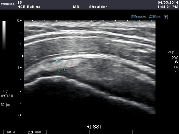
Subacromial bursitis
Image 1A Subacromial bursitis. The image demonstrates a thickened bursa which is easily diagnosed on ultrasound.
Image1B shows, using a real-time ultrasound technique, the needle placed in the exact location required. Precise needle positioning with millimetre accuracy is achievable.

Image 1B Ultrasound Image clearly showing the location of the needle into the thickened bursa.
The benefit for this patient was a significant decrease in pain and significant increase in the range of movement.
Trochanteric bursitis
This patient presented with trochanteric bursitis. There is a lot of adipose tissue to penetrate making accurate placement of the needle difficult. However, under ultrasound accurate placement of the needle was achievable.
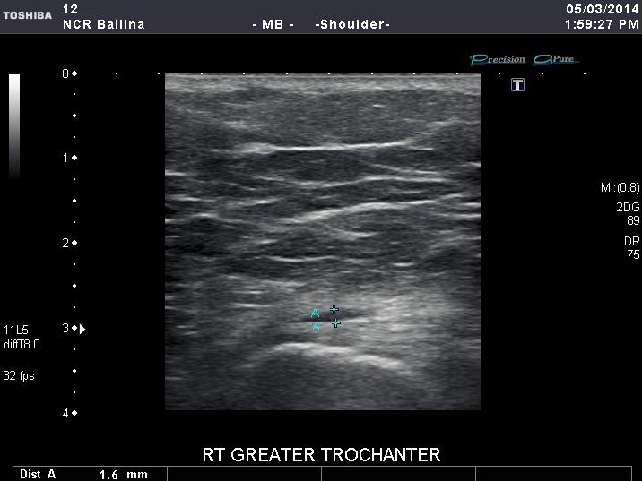
Image2A Right greater trochanter showing bursa location
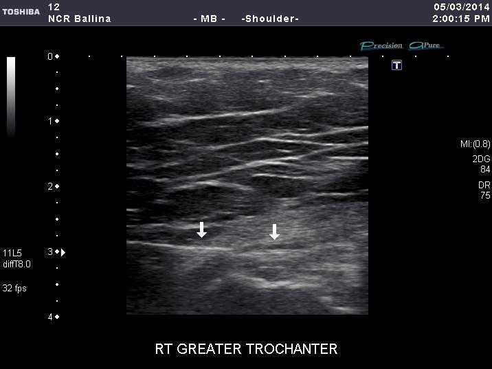
Image 2B The needle is clearly visible as it moves towards the bursa
With this level of accuracy, patients are less likely to be subject to painful repositioning. of the needle. Also the free passage of steroid and local anaesthetic around the bursa can be observed in real time. Skilled professionals are able to perform these injections easily with great accuracy . Therefore, the benefits to patient care are clear to see.
Mark Bryant
Senior Sonographer
North Coast Radiology